Following a good time at the casino the
weekend before last I thought I’d play Russian roulette with what specialty I’d
end up working in today. So I wondered in to work and got soaked in the
downpour. I could easily have stayed in bed and listened to the raindrops
hammering against the tin roof, but alas duty called!
 |
| Raining! |
Anyway as I walked
towards theatres one of the trainees who was leaving after the night shift of
the weekend informed me of the desperately sad news that a lady that I had been
a part of looking after had passed away on the intensive care unit overnight.
Sadder still, as she came to hospital in labour with her first child. Amongst
the emergency casearian section for shoulder dystocia the baby didn’t make it.
She bled and was stabilized with a hyseterectomy and moved to the intensive
care unit only to bleed for the second time the following day (when I got
involved in the case) and did remarkably well until last night when her kidneys
that had been failing stopped working over the weekend and her lungs filled
with fluid and she eventually passed away. Tremendously sad story and leaves a
husband and family bereft. Incredibly sad and frustrating that this continues
to happen despite all the courses and the advancement of medicine here. But it makes working here all the more
important. So that shook off my Monday apathy and I strode purposefully to
theatre.
When I got to main theatres it was a pleasant surprise that there were a
few hands on deck so to speak. And the ship seemed to be sailing in the right direction
for once! So whilst looking around at who was where and what was going on I
happened across a few interesting cases. Now to many medics in the UK – when
one mentions that one is going to do a ‘lumps and bumps’ list – it generally
means a few little excisions of cysts, lipoma’s or abscesses or sometimes even
a hernia is counted in this. Basically small fry operations (not to the patient
or surgeon of course).
However here, lumps and bumps take on a
whole new meaning! And not just any type of lumps and bumps – but neurosurgical
(brain surgery!) lumps and bumps.
We started with a patient who had some of
the largest collections of lumps I have ever seen. They had a condition called
von Recklinghausen’s or Neurofibromatosis. These cause loads of lumps and brown
skin stains (café au lait patches) all over the skin. This patient has a textbook
entry of them all over her body but had come as there was the largest
neurofibroma lump I have ever witnessed covering her hip and thigh. So large
that she had trouble doing up her Chitengi (which is not unlike a sarong skirt)
– which was the reason she presented for surgery. However this passed without
too much of a hitch and she will soon be able to go about her normal business
wearing her normal clothing.
 |
| Cafe au lait spot and a neurofibromatosis on back of knee |
The second had two of the largest bumps I
have ever seen growing out of a skull. To begin with we thought they were just
fatty tissue under the skin lipoma) but on looking at the x-ray we could see
that they’d eroded through some of the skull. In fact the neurosurgeon decided
to do what the red Indians did – a scalping in order to get at them easier
(under anaesthetic of course!). Anyway it turns out that these were not lipomas
or cysts but they decided that they were secondary metastatic cancer spread
from somewhere else as they extended into the skull. I have my doubts that this
is the correct diagnosis but then again these are the first lot of these bumps
that I have ever witnessed so it may well be true…. I hope to find out before I
leave!
 |
| 2 large bumps |
So
having warmed up nicely (well actually with the new aircon blasting down on us
it was rather cold) the surgeons decided to press ahead with the first patient
on the list. A brain tumour. A meningioma to be precise and a very large one at
that in a chap that is a year younger than myself (yes that does make him 20!
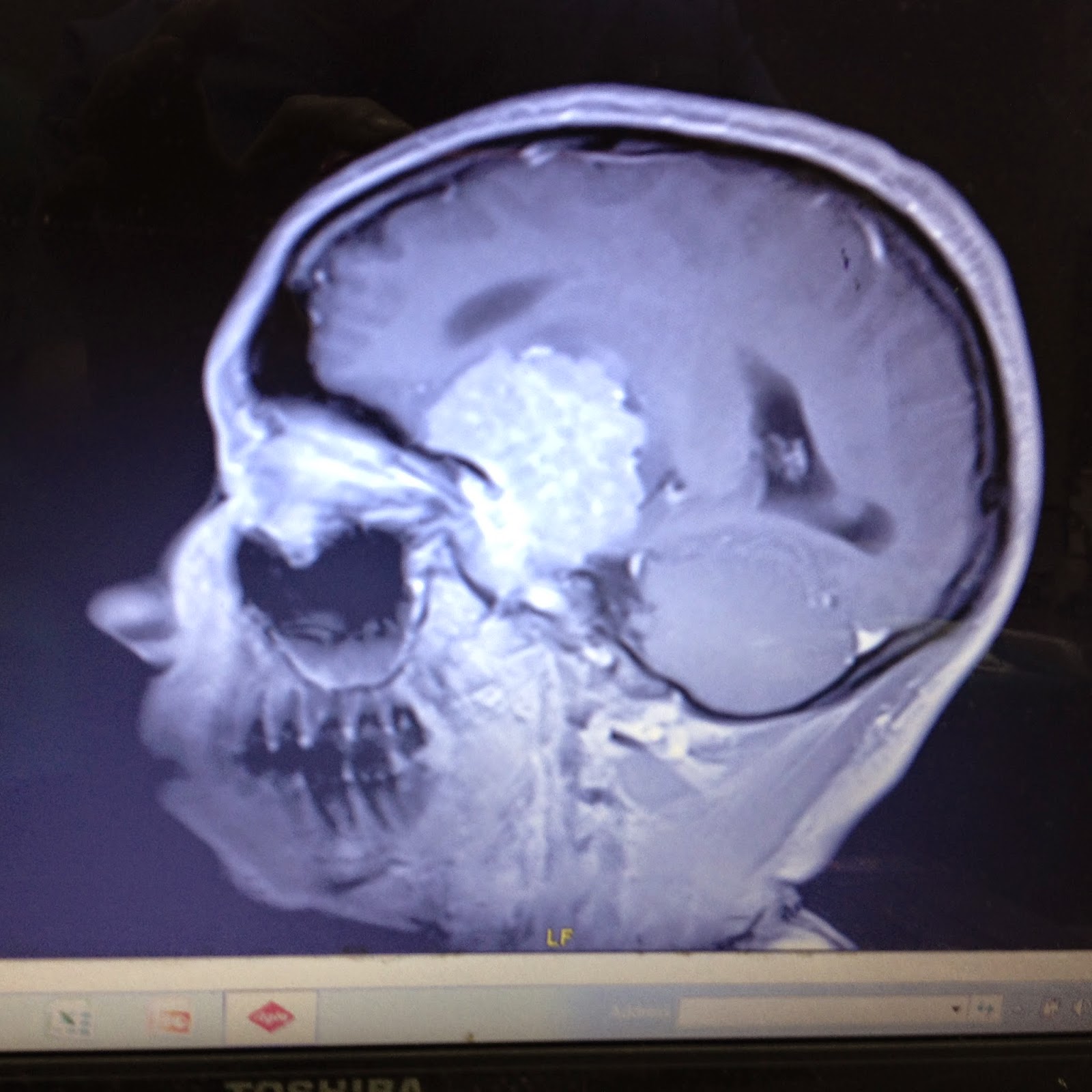
Ahem!). The scan is remarkable for two reasons – firstly its size and they are
operating with no clever technology (stealth) but also that it is an MRI scan
not a CT. Now anybody who’s a part of the NHS or even had a scan knows how long
they had to wait to get a CT scan let alone an MRI scan – so how on earth have
we got them for a brain tumour…. Well, it seems when the CT scanner in the main
university hospital in Zambia hasn’t worked for about 4 years, and the back up
CT scanner across the car park at the Cancer diseases hospital has also stopped
working as of 3 months and the Military hospital cannot deal with the now
increased influx of CT scans (even being taken from our ICU in an ‘ambulance’ –
well a van with a cylinder or oxygen in the back) then all these cases will get
an MRI scan at the cancer disease hospital instead (the MRI in UTH is of
course… out of action!)
 |
| Massive meningioma |
Anyway at 12.30 a craniotomy for meningioma
(basically taking the skull off to shell out the tumor) is not what you want to
hear, bearing in mind that the standard operating times in Zambia is until
13.30…. It was going to be a long one…. It was also going to clash with the
afternoon critical incidents meeting and teaching session that we’d organized.
However there was a need to do the case and so we proceeded.
We
started off with the pleasing aspect of getting everybody in theatre doing the
WHO Surgical Safety Checklist as led by one of the anaesthetic trainees. This
is to prevent operating on the wrong person/site or missing vital information
such as allergies or problems and also encourages good communication and teamwork.
It isn’t used well everywhere but the neurosurgeons are one of the better
surgical disciplines at doing it.
 |
| Mmed trainee leading WHO Checklist |
Then
the surgeons buried themselves in their work and I dashed out to the ‘canteen’
to get food. I usually take in a packed lunch but this morning in my lazy state
I decided I’d not bother. Whilst standing in a long queue to pay for some food
I then recalled why I usually bring my own. Nevertheless I got to the front
only to be told I had stood in the wrong queue and this one was for doughnuts
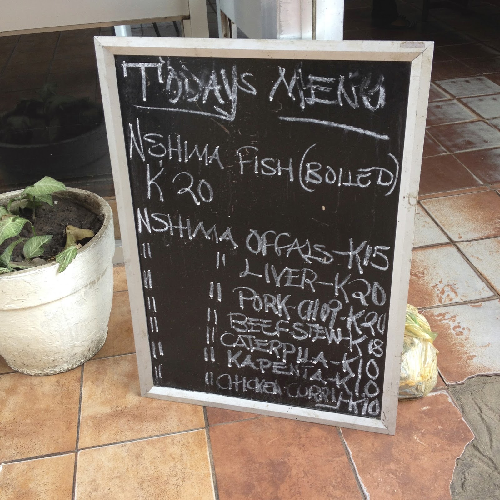
and scones. So I decamped to the other one where I paid for my Chicken and rice
(not brave enough to have caterpillars in the hospital – have tried at a
restaurant previously!). Anyway 20 minutes later as I get to the front of this
queue there is nearly a revolt as they run out of chicken ...Seriously, the
only thing that could have been worse would have been the news of no more
Nshima – so furious were the crowd! Thankfully thought it was a
misunderstanding and they just required re-stocking of the chicken and order
was restored!
I
get back to theatre and think its odd as my colleague Papari and Dave were both
missing. It turns out that whilst I was desperately trying to secure some lunch
they were desperately trying to resuscitate an emergency on intensive care
(which is next to the main theatres). Turns out it was my trauma patient from a
week ago (the one with a fractured pelvis and no chest x-ray who later turned
out to have a fractured neck, fractured ribs and haemopneumothorax blood and
air outside his lung), fractured lumbar spine and pelvis). He had also
developed kidney failure from rhabdomyolysis (muscle breakdown) over the
weekend. He also sadly didn’t make it despite their best efforts. Sadly another
frustratingly young death to be involved with.
 |
| DIY Drill |
 |
| Hand saw / cheese wire drill |
By
the time I got back and look closer at what the surgeons were doing it seems
that they hadn’t actually removed the skull at this point in time. It seemed
inordinately long to me but the flip side was that the blood loss was very
minimal (just as well as we only had 2 units of blood available anyway!) They
then broke out the ‘brain drill’. This sounds like some serious piece of kit.
It is sadly however a basic Black and Decker drill (a la what you use on a Bank
Holiday Monday to hang pictures on the wall!) wrapped in a sterile drape.
Thankfully today the batteries were charged and it made the requisite 5 holes
it needed. They then painstakingly hand saw the bones between the drill holes
to remove the bone flap. This was
interesting to watch at least as it was something that I could actually see and
not just the constant digging around in the head that continued for the next 4
hours after this!
 |
| 2 anaesthetic charts stuck together is a sign of a long operation! |
They
got as much tumour as they could out. At some stage one of the porters arrived
with quite a few polystyrene boxes of chicken and chips and a bright orange
(teeth dissolvingly sweet) fizzy drink called Miranda. Apparently if the staffs
stay past 13.00 then the surgeon/hospital is bound to provide them with lunch.
So credit where credits due the surgeon did look after his staff – thought the
poor scrub nurse’s one was stone cold by the time the operation finished! It
also meant I could have avoided the hour of my life at the canteen queue
system! Every day’s a school day I guess!
 Anyway after a painful/painstaking (depends on
whether you are looking from an anaesthetic or surgical perspective!) 6 and a
half hours they decided they’d got as much tumour out as they could today. On
further clarification I was told that they weren’t going to get the rest and
anyway it would grow back in due course!!! 6.5hours later!!!! So we took him
over to ICU where we woke him up and he’s ok if not a little groggy after all
that Halothane! I am crossing every fibre in my body that this remains the case
overnight, as my current ICU statistics are looking a little grim. Though not as bad as the realisation that my own oxygen saturations are way lower than the patients - even on ICU at times! Well.... it was a long case, I had to find something to do!!
No comments:
Post a Comment